CASE REPORT
SAMPAIO, Amanda Talys [1], MARQUES, Daniel de Lima Fermino [2], SCUDELLER, Melissa Ziglio [3], GIOMETTI, Ines Cristina [4], CASTILHO, Caliê [5]
SAMPAIO, Amanda Talys et al. Bilateral polycystic ovary in a nellore cow: case report. Revista Científica Multidisciplinar Núcleo do Conhecimento. Year. 09, Ed. 12, Vol. 01, pp. 05-11. December 2024. ISSN: 2448-0959, Access link: https://www.nucleodoconhecimento.com.br/veterinaria-en/bilateral-polycystic-ovary, DOI: 10.32749/nucleodoconhecimento.com.br/veterinaria-en/bilateral-polycystic-ovary
ABSTRACT
The most common ovarian disease in cattle is follicular cyst, but there are few studies addressing this problem in zebuid cows on pasture. In humans, this condition is known as polycystic ovary syndrome (PCOS). The aim of this study is to report on the occurrence of polycystic ovary syndrome in a multivariate Nellore cow kept on pasture. The case was detected during an ultrasound examination of the reproductive tract at the beginning of the breeding season. The cow in question was approximately 60 days postpartum and rectal palpation revealed bilateral ovarian enlargement. Ultrasound examination revealed a low-echo lobular image suggestive of multiple follicles. Without treatment, the cow was slaughtered and her reproductive tract removed for macroscopic, morphometric and histopathologic analysis. The ovaries were found to contain a large number of cystic formations of varying sizes, confirming the diagnosis of polycystic ovary syndrome. Although PCOS is well documented in humans, it has not yet been described in female cattle. This report is novel in that cows kept on pasture and fed only mineral salt rarely show ovarian pathologies. It can therefore serve as a guide for professionals in the field who are aware of the possible occurrence of this case.
Keyword: Ovarian pathology, Zebu cattle, Follicular cyst.
1. INTRODUCTION
Among the diseases of the ovaries, the follicular cyst is the most common in female cattle. It is characterized by the presence of a fluid-filled structure in the ovaries for at least ten days with a diameter of more than 2.5 cm, which for some reason does not induce ovulation and thus prevents the normal cyclicity of the female1,2.
This condition is more frequent in dairy cows because they are fed a nutrient-rich diet3. Zebu cattle have a lower incidence of follicular cysts2. In humans, this disorder is known as polycystic ovary syndrome (PCOS)4 Like women, cattle are monovulatory, i.e. they usually have only one dominant follicle that releases a single oocyte in response to LH5.
The aim of this study is to report on the occurrence of polycystic ovaries in a multiparous Nellore cow kept on pasture.
2. CASE HISTORY
The case occurred at the UNOESTE experimental farm in the municipality of Presidente Bernardes, São Paulo, Brazil, at 22º 28’ 25” S, 51º 67’ 88” W and an altitude of 400 meters. The climate is tropical, with an average annual temperature between 15ºC and 32ºC. Diagnosis of the cows’ reproductive tract was carried out using ultrasound before the start of the breeding season. The cows are kept in the Panicum maximum cv. Tanzânia pasture with free access to water and mineral salt.
The gynecological examination was carried out in January 2022 on a 4-year-old perennial Nellore cow. The interval between calvings of the last pregnancy was 352 days, with the first calving on 10.09.2020 and the second on 26.09.2021. At the time of discovery of the ovarian abnormality, the cow was approximately 120 days postpartum.
Rectal palpation revealed bilateral ovarian enlargement, and ultrasonography (Honda model HS-2000VET with a 7.5 MHz transrectal transducer) revealed a lobular, anechoic image suggestive of multiple follicles greater than 7 mm in size (Figure 1).
Thirty days later, a follow-up ultrasound was performed, which showed that the ovarian condition was unchanged, with the presence of multiple follicles (Figure 2). No treatment was carried out and the decision was made to slaughter the cow. In June, the cow was slaughtered and the reproductive tract was removed immediately after slaughter and sent to the pathology laboratory for macroscopic analysis, morphometry and preparation of samples for histopathological examination.
After macroscopic evaluation with photo documentation (Figure 3), the right and left ovaries were weighed and measured and the length and width of the right and left uterine horns were recorded (Table 1). Longitudinal sectioning of the ovaries with a scalpel revealed a small hematoma-like area in the left ovary and a corpus luteum in the right ovary (Figure 4).
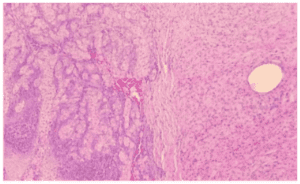
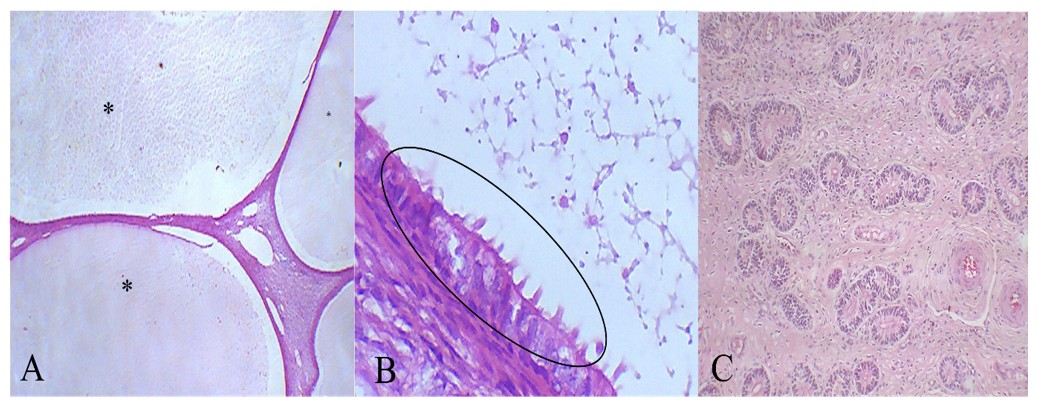
The ovarian samples were fixed in 10 % buffered formalin and subjected to a standard histological examination. Histopathologic examination revealed that the right and left ovaries had a large number of cystic formations of different sizes, with walls composed of follicular epithelium and secretory contents ranging from pink to amphophilic and diffusely distributed in the cortical and medullary regions (Figure 5).
The endometrium was also examined histopathological and showed no significant pathological changes (Figure 6). The final diagnosis was polycystic ovary syndrome.
3. DISCUSSION
This appears to be the first reported case of polycystic ovaries in a young Nellore cow kept exclusively on pasture and for which there are no predisposing factors. Cystic ovarian disease in cattle is well documented and relatively common. However, unlike the case presented here, it usually involves one or a few follicles that exceed the breed-typical ovulation diameter. We have classified our finding as a polycystic ovary because the morphologic and ultrasonographic features are similar to polycystic ovary syndrome (PCOS) in humans. PCOS is well described and studied in humans, but there are no reports of it in female cattle, especially in zebu breeds.
The first significant finding in this case was the marked bilateral enlargement of the ovaries on rectal palpation. In a study of ovaries of Zebu cattle obtained from slaughterhouses, the right ovary measured 2.73 cm, 2.04 cm and 1.71 cm and the left ovary measured 2.67 cm, 1.94 cm and 1.70 cm in terms of length, width and thickness, respectively. These measurements were larger (p<0.05) when a corpus luteum (CL) was present6. The polycystic right ovary in this case did not show the same increase in size due to the presence of a corpus luteum, as it was already abnormally large. In a recent study in which the CL was monitored from ovulation to luteolysis in non-pregnant and non-supplemented Nellore heifers, the CL reached a maximum diameter of 1.93 cm on day 12 of the estrus cycle7. In the present case, the CL was measured at 1.78 cm during an unknown phase of the estrous cycle, which is within the normal range. In humans, according to the Rotterdam8 criteria for PCOS, pelvic ultrasound shows at least 12 follicles in each ovary ranging in size from 0.2 to 0.9 cm and/or an increase in ovarian volume without the presence of a dominant follicle or corpus luteum.
Follicular cysts in cattle typically occur during the first postpartum cycle and are associated with anoestrus, potentially recurring over several consecutive cycles and leading to economic losses9, 10. It has been reported that in dairy cattle breeds, the most common symptom of cysts is anoestrus, with disease incidence being highest up to 90 days after parturition3. In the present study, the zebu beef cow was approximately 120 days postpartum, had multiple cysts and was pregnant due to the presence of a CL.
A histologic examination of the endometrium was performed because it showed hypertrophy macroscopically; however, no significant pathologic changes were found. Other ovarian pathologies can also occur in female cattle, e.g. granulosa cell tumor, abscesses, dermoid cysts, teratomas and rete ovarii metaplasia2, so histopathologic examination is required for a reliable diagnosis .
This case report is a novelty as cows kept on pasture, fed only mineral salt and young rarely present ovarian pathologies, especially polycystic ovary disease, the occurrence of which has not been documented in cows. This finding in a routine ultrasound examination serves as a guideline for professionals in the field regarding the possible occurrence of this type of cystic disease in female Zebu cattle.
REFERENCES
- Grunert E, Birgel EH, Vale WG & Birgel Júnior EH. Distúrbios da Reprodução dos Animais Mamíferos Domésticos com Sede nos Ovários. In: Patologia e clínica da reprodução dos animais mamíferos domésticos: Ginecologia. 1th ed. Virela, São Paulo, Brazil, 2005, pp. 316-318.
- Nascimento EF, Santos RL. Patologia do Ovário. In: Patologia da Reprodução dos Animais Domésticos. 4th ed. Grupo GEN, Barueri, Brazil, 2021, pp. 24-25.
- Fernandes CAC, Figueiredo ACS, Oba E, Viana JHM. Factors Predisposing for Ovarian Cysts in Holstein Cow. ARS Veterinária. 2005; 21(2): 287-295.
- Azziz R. Polycystic Ovary Syndrome. Obstet Gynecol. 2018; 132(2): 321-336. https://doi.org/10.1097/AOG.0000000000002698
- Ginther OJ, Wiltbank MC, Fricke PM, Gibbons JR, Kot K. Selection of the dominant follicle in cattle. Biology of Reproduction. 1996; 55:1187-94. https://doi.org/10.1095/biolreprod55.6.1187
- Sakate NM, Guaberto LM, Giometti IC, Castilho C. Morfometria ovariana e a qualidade dos o ócitos de vacas zebuínas abatidas. Revista Acadêmica Ciência Agrária Ambiental. 2013; 11(3): 223-228. https://doi.org/10.7213/academica.011.003.AO01
- Mattos GM, Ropelli BM, Membrive CMB, Baltazer AL, Souza LFA, et al. Corpus luteum morphology and function after timed artificial insemination (Tai) in Nellore heifers supplemented with sunflower seed in their diets. American Journal of Animal and Veterinary Sciences. 2021; 16(3): 121-127. https://doi.org/10.3844/ajavsp.2021.121.127
- Rotterdam ESHRE/ASRM‐Sponsored PCOS consensus workshop group. Revised 2003 consensus on diagnostic criteria and long‐term health risks related to polycystic ovary syndrome (PCOS). Human Reproduction. 2004; 19(1):41–47. https://doi.org/10.1093/humrep/deh098
- Ortega HH, Marelli BE, Rey F, Amweg AN, Díaz PU, Stangaferro ML, Salvetti NR. Molecular aspects of bovine cystic ovarian disease pathogenesis. Reproduction. 2015; 149(6):R251-64. https://dx.doi.org/10.1530/REP-14-0618
- Gupta C, Kumar S, Murugan M, Ramprabhu R. Management of Follicular Cyst by Multiple Approaches in Crossbred Cows. International Journal of Livestock Research. 2020; 10(10):164-168. http://dx.doi.org/10.5455/ijlr.20200507083031
Table 1. Morphometry of ovaries and uterine horn of Nellore cow with polycystic ovary
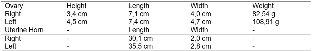
Figure 1. A – Ultrasound in January 2022. B – Ultrasound in February 2022; C – Whole ovaries along with the uterus; D – Whole ovaries. E – Sectioned ovaries; F – Sectioned left ovary of a Nellore cow with polycystic ovary; G – Endometrium (black arrow) and cervix (white arrow); H – Left ovary with a hematoma. I – Right ovary with the presence of a corpus luteum, obtained from a Nellore cow with polycystic ovary
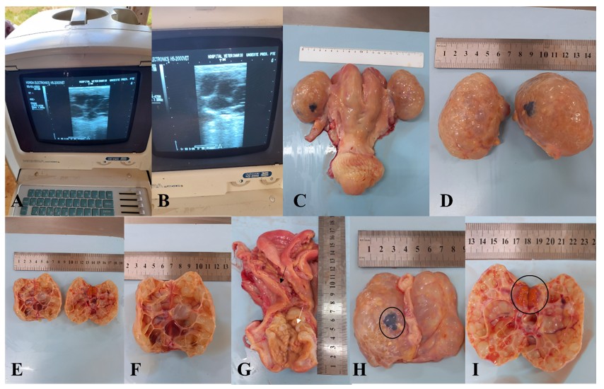
Figure 2. Histological photomicrograph of the ovaries of a Nellore cow with polycystic ovary. A – Extensive cystic formations, follicles with amorphous amphophilic secretory material (*). H&E stain, 10x objective. B – Highlighted follicular epithelium. H&E stain, 40x objective. C – Absence of significant pathological alterations in the endometrium. H&E stain, 10x objective
[1] Graduated in Veterinary Medicine from the University of Western São Paulo. ORCID: https://orcid.org/0009-0003-0962-6173. Currículo Lattes: http://lattes.cnpq.br/6704212946922532.
[2] Graduated in Veterinary Medicine from the University of Western São Paulo. ORCID: 0009-0008-9759-4824.
[3] Graduated in Veterinary Medicine from the University of Western São Paulo. ORCID: https://orcid.org/0009-0003-3076-7536. Currículo Lattes: http://lattes.cnpq.br/7320103488939692.
[4] Post-doctorate at the University of São Paulo (USP); PhD in Veterinary Medicine [Botucatu] from the São Paulo State University Júlio de Mesquita Filho; Master’s degree in Veterinary Medicine from the Federal University of Santa Maria; Degree in Veterinary Medicine from the University of Western São Paulo. ORCID: https://orcid.org/0000-0001-8621-8374. Currículo Lattes: http://lattes.cnpq.br/5461216137094368.
[5] Supervisor. PhD in Veterinary Medicine from the São Paulo State University, Jaboticabal Campus; Master’s Degree in Biological Sciences (Pharmacology) from the São Paulo State University, Botucatu Campus; Residency in Animal Reproduction from the São Paulo State University, Botucatu Campus; Degree in Veterinary Medicine from the University of Western São Paulo. ORCID: https://orcid.org/0000-0003-3300-8116. Currículo Lattes: http://lattes.cnpq.br/4860674666647521.
Material received: October 9, 2024.
Material approved by peers: October 21, 2024.
Edited material approved by authors: December 12, 2024.