ORIGINAL ARTICLE
DIAS JUNIOR, Julio Cesar [1], SILVA, Fransérgio da [2], TANCLER, Murilo Colino [3]
DIAS JUNIOR, Julio Cesar. SILVA, Fransérgio da. TANCLER, Murilo Colino. Occurrence of asymmetries of lower limbs in basic futsal athletes. Revista Científica Multidisciplinar Núcleo do Conhecimento. Year 06, Ed. 01, Vol. 05, pp. 05-29. January 2021. ISSN: 2448-0959, Access Link: https://www.nucleodoconhecimento.com.br/health/basic-futsal, DOI: 10.32749/nucleodoconhecimento.com.br/health/basic-futsal
SUMMARY
Futsal is a sport on the rise worldwide, attracting more and more new practitioners and as well as in field soccer, it has undergone changes in recent years, increasingly demanding athletes, becoming a high impact sport, promoting overload, in the short, medium and long term, predisposing to injury to different degrees of the locomotor apparatus. The aim of this study was to analyze lower limbs asymmetries in futsal-based athletes, as well as their relationship to the incidence of injuries. The study was developed with 47 athletes of the basic category, futsal, from a city in the interior of the state of São Paulo, where functional tests were performed with the help of the PHAST application. A pattern of similarity was identified between the muscle groups tested, except for the gluteus muscle, presenting a significant difference. According to the indicators found, the patterns of strength deficit of the middle glutes may trigger, or predispose to, some types of biomechanical lesions whether proximal, in the hip region, or distal, as dysfunctions in the knee joint. When an athlete presents with a weakness of this muscle, ipsilateral femur adduction, increased medial rotation and fall of the pelvis against lateral, promoting increased dynamic angle of the knee, increasing the overload in this joint. Thus, it is concluded that pre-season evaluation is important to identify possible asymmetries, implementing preventive work to correct them, in order to minimize the risks of injuries.
Keywords: Functional Assessment, Futsal Injury, Injury Prevention, Futsal.
1. INTRODUCTION
Futsal is a sport on the rise around the world, attracting more and more new practitioners (GAYARDO et al., 2012). Because of its ease of finding space for its realization, it is one of the most widespread modalities in Brazil, played by more than 12 million people (RIBEIRO and COSTA, 2006). With its proliferation, at the end of the 1980s, indoor soccer was consolidated and acquired an importance among the world population (TRIQUES, 2005; REZER and SAAD, 2005).
It is a sport similar to field soccer, being played on specific courts, called all-rounder, which are also demarcated for other sports, such as volleyball and basketball. Two teams, with five players, one ball with smaller dimensions and heavier than traditional football (KURATA et al., 2007).
As in field soccer, futsal has been undergoing changes in recent years, increasingly demanding athletes, becoming a high-impact sport (GOMES and SILVA, 2002). It is disputed by both sexes: male and female, where it has been gaining more and more space in the sports scene, and also, struggling to become an Olympic program, being paramount for the strengthening of this category (GAYARDO et al., 2012).
The evolution of futsal is related to the tactical, technical and physical aspects, increasingly investigating the performance of athletes. Thus, with the increase in the number of competitions, there is a growth in the number of games and trainings, requiring more of the human body, promoting overload, in the short, medium and long term, predisposing the injury of different degrees of the locomotor apparatus (SIMÕES, 2003; ABRAHÃO et al, 2009). In addition, the high-performance sport promotes high exposure, making the athlete work at his limit of physical, psychological and mental endurance, resulting in these injuries, such as muscle injuries (OLIVEIRA, 2007).
It is a modality that requires acceleration and deceleration movements with sudden changes of directions, exposing its participants to excess impacts, triggering musculoskeletal and oxidative damage (SOUZA et al., 2010).
The literature states that, when adolescents start soccer practice, intense training, bodybuilding and little stretching, develop an athlete with little flexibility, consequently hindering the sports gesture (kick) with little precision and less power, due to the flexibility deficit of the hamstrings (GRAU, 2003). This muscle (posterior thigh), together with gastrocnemius are more prone to stretching, because they are biarticular and due to eccentric overload can trigger muscle injuries: hamstrings in kick deceleration; gastrocnemius on landing (COHEN and ABDALLA, 2003; BERTOLA et al., 2007). This muscle tissue injury corresponds to 20 to 40% of sports injuries, obtaining a predominance of 80 to 90% in the lower limbs (JUNIOR, 2019).
These eccentric movements trigger increased creatine kinase (CK) levels in the bloodstream, directly linked to the generation of free radicals. In addition, the increase in oxygen consumption during exercise promoting the increase in the production of reactive oxygen species, contributing to muscle injuries and inflammatory processes (SOUZA et al., 2010; CRUZAT et al., 2007). In this case, physical training alters the balance, and can balance the pro and antioxidant agents, depending on the frequency, intensity and duration of the specific activity (PINHO et al., 2006).
These injuries resulting from the practice of futsal and the risks to its events have become a focus of concern for health professionals, after all, they leave athletes away and disabled for various periods of training and competitions to be rehabilitated correctly (GAYARDO et al., 2012). There are modern features of electro, kinesiotherapy and manual therapy, which accelerate the return process, since competition requires a quick return to the courts (KURATA et al., 2007).
However, etiologies can be numerous, from direct (trauma) or indirect injuries (repetitive overload) and/or degenerative disorders, since high-level sport requires issues of muscle strength, range of motion and weight transfers far above the physiological, such as: running; jumps; starters; heads among others (FRANCA et al., 2004; DIAS JUNIOR et al. 2018). Thus, when these issues are exacerbated by unbalancing the system of strength, resistance and flexibility, there is an accumulation of musculoskeletal and myotendinous stress triggering the problem, the neuromuscular pattern suffers a collapse, influencing physical quality, decreasing performance, adhesions and fibrosis in the fascia, reducing tissue fluidity, presenting pain, edema, ischemia, muscle tension, spasms (SANDANDOVAL, 2005; DIAS JUNIOR, 2020).
In addition to futsal being practiced professionally, there is a large number of recreational practitioners, which becomes a problem, because it does not have a work periodization, specific training, systematized, associating with low physical conditioning, overweight and individual characteristics of each athlete, favoring the risk of injuries. Therefore, it is extremely important to evaluate the characteristics of each individual and correlate with the main lesions, seeking methodologies that can influence and reduce the number of injuries, improving the effects of health and quality of life of practitioners (PINHEIRO and ROCHA, 2017).
In rehabilitation it is important to restore neuromuscular function, improve strength, proprioception, cardiorespiratory capacity and recovery of the sports gesture. In addition, to enhance the maximum functions of the athletes, guidance on training, maintaining static and dynamic postures, preventing recurrences of injuries and readapting the individual to the practice of the sport (KURATA et al., 2007). For this it is necessary to investigate the incidence, risk factors, mechanism of injury, filing appropriate preventive strategies, aiming at prophylaxis and percentage reduction of injury (VANDERLEI et al., 2010; DIAS JUNIOR et al., 2018).
With all these specifications that happen with the practice of futsal, it is established that physical body conditions are extremely important for the performance and prevention of injuries. However, this study seeks to evaluate the occurrence of lower limbs asymmetries in futsal athletes.
2. GOAL
To analyze lower limbs asymmetries in futsal-based athletes, as well as their relationship to the incidence of injuries.
3. METHODOLOGY
The present study was submitted to the Ethics and Research Committee of the University of Araraquara, through the Brazil platform, approved with the opinion number: 4,419,631. The athletes were informed about the objective and design of the present study, as determined by Resolution 466/12 of the National Health Council and Circular Letter 166/18 of the Ministry of Health. Therefore, they agreed to participate and signed the free and informed consent form and parents and legal guardians signed the free and informed consent form.
The research was conducted with athletes of the basic category, futsal, from a city in the interior of the state of São Paulo, participating in the sample, 47 athletes aged 8 to 18 years, who competed in championships of this modality and maintained a weekly training schedule of 4 hours.
The athletes were invited to participate in the research by the physiotherapist in charge of the team, who is a participant in this research. The terms of nods for each athlete and also the consent form for the parents and legal guardians of each player were presented and explained.
The evaluations occurred through specific functional tests: muscle strength of hip extensors; range of motion (ROM) of ankle dorsiflexion; flexibility of ischiossurais; pelvic bridge; middle gluteus function; hip stiffness. All these trials were applied to the lower, bilateral limbs of the athletes, using a support tool, called PHAST (Phisiotherapy Assessment Tool), which corresponds to a functional evaluation application, performed by the physiotherapists responsible for this research.
The athletes presented themselves at the club, in the morning, in November 2020, after the approval of the research by the ethics committee, being at rest and without performing any type of physical or technical intervention before the evaluation. Therefore, they attended the club before the time of the respective training sessions.
The sequence of the PHAST evaluation protocol was followed, where personal and anthropometric data were first collected: name; e-mail; phone number; sex; date of birth; leg kicking; hand writing; diet; height; weight; average sleep per night. Following was added characteristics of the activity: football; beginning of the practice; games/competitions per week; training week (hours per week); observations if necessary. He then added possible previous injuries: site of the lesion; injured limb; injured structures; mechanism of injury; type of injury; contact injury; pain level; surgery has been performed; date of injury; days of immobilization; days away; numbers of therapeutic care; observation if necessary.
Secondarily functional tests occurred in the order that the application recommended and that was mentioned earlier.
To measure angulation in the tests that required the score in degrees (range of motion – ROM of ankle dorsiflexion; flexibility of ischiossururals) an application called Angle Meter was used, installed on the smart phone of the Brand Sansung, Galax A30 S model, in which it rested on the limb of the individual and measured the inclination that the leg presented. This use of applications for slope assessment, measured in degrees is scientifically validated and widely used in clinical practice (MELQUIADES, 2018).
The values collected were recorded in the Phast application, where at the end of each evaluation presented an analysis of particular symmetries or asymmetries of each individual.
Data were collected at the end of all evaluations and transferred in a spreadsheet in the apple numbers software for data analysis.
To evaluate the difference between the asymmetries found, the Kruskal-Wallis nonparametric test (alpha= 0.05) was used. Bray-Curtis cluster analysis (UPGMA) was used to verify the similarity in the applied events, with data on the proportion of quantified asymmetries. The calculations were performed with the help of the PAST-version 4.03 software (HAMMER et al., 2001).
Description of the protocol of the specific tests:
Before starting the tests, the athletes performed 10 minutes of warm-up on an exercise bike to improve the vascularization of the lower limbs, aiming at an improvement in performance and a possible prevention of late muscle pain.
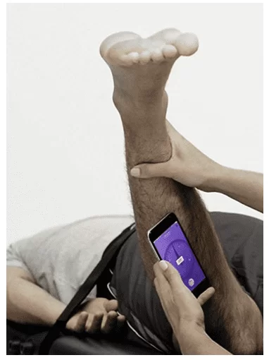
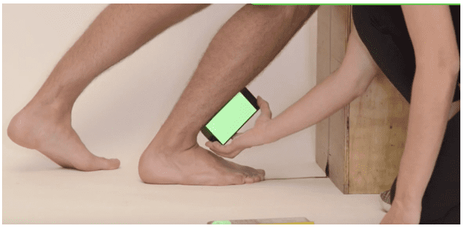
– Hip stiffness test: the athlete remained lying in ventral decubitus, relaxed, with a stabilized pelvis. The physiotherapist performed a knee flexion of the tested limb and located the anterior tuberosity of the tibia, marking 5 centimeters above hers, while the other limb was extended. With the knee in flexion at 90º, it was verified if the hamstrings were relaxed, subsequently performing an internal rotation of the hip to the limit of movement, without compensation of hip elevation or lumbar spine movement. With the help of the cell phone, in the mark that was performed on the tibia, the angulation of ROM was measured (Figure 1).
Figure 1: Test for hip stiffness.
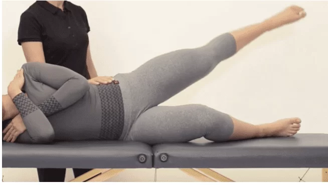
– Middle gluteus function test: the athlete lay in lateral decubitus, with the lower limb leaning against the stretcher, frailted, and with the limb to be tested upwards, free to perform hip abduction, with a slight extension. Performed contractions counting the maximum repetitions, until modifying the movement pattern or presenting compensations of other structures, such as the pelvis of the tested limb unbalance and rotate to the anterior or posterior; and/or perform knee flexion of the tested limb (Figure 2).
Figure 2: Test for middle gluteus function.
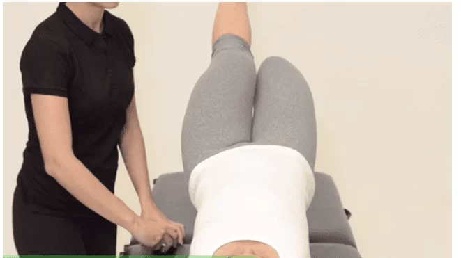
– Pelvic bridge test: the athlete remained in supine position with knee flexion. At the time of the test extended one of the knees, while the contralateral foot remains on the stretcher. Then raised his hip and asked to keep high for 10 seconds. It was observed whether at any time the misalignment of the pelvis occurred and grading as mild, moderate or severe (Figure 3). For example, the ideal is to keep the knees and pelves aligned and without compensation, misalignment of the hips, or lowering of the spine.
Figure 3: Pelvic bridge test.
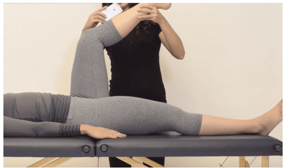
– Ischiosural flexibility test: athlete in supine position, kept an extended leg on the stretcher, while the contralateral leg, to be tested, was maintained at 90º of hip flexion, with the ischiosurals relaxed. The physiotherapist extended the knee of the tested limb until he felt a final resistance offered by the muscle. He used the cell phone to measure the degree of angulation, below the anterior tuberosity of the tibia (Figure 4). This test assesses a retraction of the ischiotial and triceps beating muscles.
Figure 4: Ischiossurial flexibility test.
– Ankle dorsiflexion ROM test: athlete was left with his foot resting on the ground near the wall. He then identified the anterior tuberosity of the tibia, marking 15 centimeters below, where the cell phone would be placed with the inclinometer. He then asked to perform the knee flexion until he touched the wall, without untouching the heel of the floor. The cell phone was used to measure the angulation by engaging in the tibia marking (Figure 5). If in the knee flexion to touch the patella on the wall, the athlete pulls the heel off the floor, ask the player to zoom in a little more from the foot of the wall, until he can touch the knee against the wall without touching the heel.
Figure 5: Test for ankle dorsiflexion ROM.
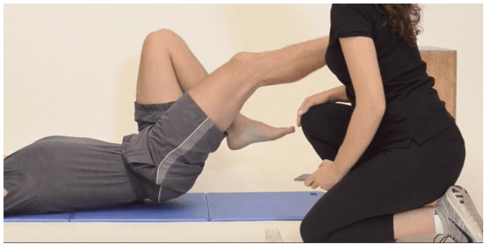
– Test of muscle function of the hip extensors: the athlete lying on the mattress, with his hands behind his head and supported one of the heels in a chair or bench 60 centimeters high. Using a goniometer, the knee of the tested limb was measured and maintained at 20º, while the contralateral limb was in the position of 90º hip and knee flexion. He then asked the player to perform the contraction of the muscles, raising the hip from the ground, counting the maximum repetitions, until presenting some type of compensation, such as: starting to move the untested limb (leaving 90º) in an attempt to increase the strength of the tested limb, or increase the speed of movement to compensate for the force deficit, reducing the hip extension ROM and reducing the 20º knee flexion (tested figure 6).
Figure 6: Test for muscle function of hip extensors.
4. RESULTS
The results found are presented in graph 1, boxsplot, which shows the distribution of the data, apparently meaning that the data have a similar distribution in terms of the types of imbalances, for example, the ischiossural is much more concentrated than those of the middle gluteus, but the tendency is that they have the same behavior.
Figure 1: Presentation of the types of imbalances.
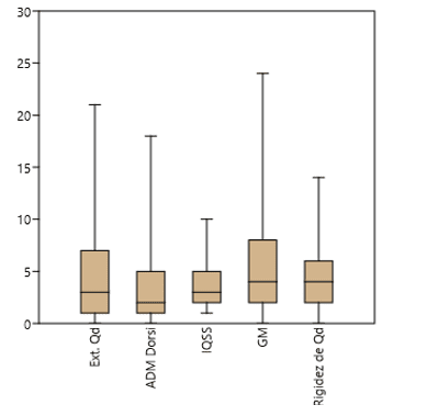
Table 1 is a descriptive analysis, basic statistics, presenting minimum, maximum, sum and mean number of imbalances, where the middle glute presented a higher mean alteration between the lower limbs. The other tests have the same constancy of imbalances.
Table 1: Analysis of basic statistics of imbalances.
| Hip extenders | ADM ankle dorsiflexion | Hamstrings | Middle gluteus | Hip stiffness | |
| Number | 47 | 47 | 47 | 47 | 47 |
| Minimum | 0 | 0 | 1 | 0 | 0 |
| Maximum | 21 | 18 | 10 | 24 | 14 |
| Sum | 207 | 164 | 163 | 292 | 207 |
| Average | 4,404255 | 3,489362 | 3,468085 | 6,212766 | 4,404255 |
| Variance | 15,81129 | 10,55967 | 5,471785 | 33,17114 | 8,680851 |
| Standard deviation | 3,976341 | 3,249564 | 2,339185 | 5,759439 | 2,946328 |
Source: Author
From the statistical point of view, when comparing all tests, a Kruskal-Wallis analysis (alpha= 0.05) was performed, where the result was close to the critical value, p= 0.53, meaning that they tend to be similar, but, as it is close to the critic, it may be that the average gluteus asymmetry is different from the others, that is, this imbalance probably presents a different median from the other imbalances (Table 2).
Table 2: Kruskal-Wallis test for equal medians.
| H (chi2): | 9,18 |
| Hc (tie corrected): | 9,32 |
| p (same): | 0,05358 |
Source: Author
When analyzing table 3, Mann whit, although the median differences are not significant, the middle gluteus has a stark difference from the others.
Table 3: Mann whit
| Hip extender | ADM Dorsiflexion | Isquitibial | Middle gluteus | Hip stiffness | |
| Hip extender | 0,2625 | 0,4228 | 0,1465 | 0,629 | |
| ADM Dorsiflexion | 0,2625 | 0,5811 | 0,01081 | 0,05151 | |
| Ischiotibial | 0,4228 | 0,5811 | 0,02444 | 0,118 | |
| Middle gluteus | 0,1465 | 0,01081 | 0,02444 | 0,3573 | |
| Hip stiffness | 0,629 | 0,05151 | 0,118 | 0,3573 |
Source: Author
Cluster analysis showed clear groups, the first group of hip stiffness being close to the ischiosural imbalance. The second relationship is of the groups, ischiosural/hip stiffness, with the adm group of ankle dorsiflexion. At the third point, the relationship of the three groups with the hip extension and finally, the group of the middle gluteus separated from all with a Euclidean distance of 45 that is quite distant (Figure 2).
Figure 2: Cluster presenting the level of similarity between the variables (cophenetic correlation = 0.97).
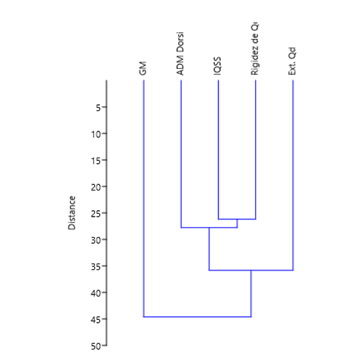
The cophenetic correlation was 0.97, presenting the stress of the analysis, that is, the cluster presented 97% of the data variation, being a very low stress, showing the behavior of imbalances in relation to body functional structures. Some imbalances are closer and others are more distant, as is the case of the group of the middle gluteus, which is distant from all groups. The ischiossural group and hip stiffness are very close.
5. DISCUSSION
Football is the sport that encompasses a large number of practitioners, with all age groups of both genders, being about 200,000 professional athletes and 240 million amateurs, 80% of which are male. It presents a lot of physical contact and a large rate of lesions (PEDRINELLI et al., 2011; ZANELLA et al., 2019).
This modality is marked by short, fast and discontinuous movements, imposing asymmetric loads, being favorable to imbalances of forces in the lower limbs, making athletes more vulnerable to injuries, where it is essential a pre-season evaluation and periodic monitoring of training to assist in prevention programs, aiming at improving athletic performance, alleviating recurrences of injury and the negative consequences for the club and the athlete himself (LEONARDI et al. , 2012; CARVALHAIS et al., 2013).
According to the data found, the results compiled in this research present a similarity between the tested groups: ischiosurals, hip stiffness, are close to dorsiflexion ROM, presenting a degree similar to that of hip extensors. However, distating from these, the middle gluteus is what is sharply distant, presenting a large discrepancy of its muscular strength, compared to the other groups tested.
Therefore, these patterns of strength deficit of the middle glutes may trigger, or predispose to, some types of biomechanical lesions whether proximal ly in the hip region, or dictated, as dysfunctions in the knee joint. Like Nyland et al., 2004, through electroneuromyography, presented a low activation of the medial vastus and middle gluteus, in hips with increased pelvic antiversion, a factor that increases the dynamic range of the knee.
When an athlete has a weakness of the middle gluteus muscle, ipsilateral femur adduction, increased medial rotation and fall of the pelvis against lateral, as he is an important hip abductor. This will cause an accentuation of the dynamic range of the knee and reduction of the contact zone of the patellofemoral joint (ZANELLA et al., 2019; FUKUDA et al., 2012). This middle gluteus dysfunction, with increased adduction and medial rotation of the hip, will trigger: when the knee is in extension – patellar laterality, causing an excessive compression of the patellar lateral face on the lateral femoral condyle; in flexion – increased load on the lateral aspect of the intercondilar femoral fossa, and may develop the patellofemoral painful syndrome (POWERS, 2003; BALDON et al., 2011; MORAIS and FARIA, 2017; GENTIL, 2018).
With this it will lead to an increase in the Q angle, influenced by three movements: tibial rotation; femoral rotation; knee vain. The vain is resulting from the adduction of the femur, leading the patella to medial in relation to the anterosuperior iliac spine. With this increase in angle will produce a lateral force under the patella, changing the alignment and causing an overload in the patellofemoral. Several factors can lead to increased vaver, they are: ligament laxity; muscle weakness in any lower limb group; genetic predisposition; high body mass index; previous lesions (ZANELLA et al., 2019; GENTIL, 2018). However Jensen and Cabral, 2006, presents that the valgos knees are not always indicative of higher value at the Q angle, conflicting with Powers, 2003, which translates biomechanical changes, such as excessive external rotation, internal rotation of the femur and valgo knee, as indicators of direct change in the value of the Q angle.
Another important factor in relation to the dynamic valve are the kinematic changes of the trunk, pelvis and hip, related to the contraction force of the muscles of the pelvic loin complex and hyperpronators of the subtalar joint developing excessive pronation in the load response phase in gait, increasing the eversion of the calcaneus, leading to medial rotation of the talusus and tibia, developing an unbalanced gait and attempting to reach full knee extension , leads to a medial rotation of the femur causing the vain (BOLING et al., 2009; ALMEIDA, 2013). Hetsroni et al., 2006, and Noehren et al., 2012, they did not present a relationship that supported the hypothesis of patellofemoral pain syndrome with the alteration of excessive pronation of the foot.
This excess of vaver disarranges the alignment of the knee, for example, in the vertical jump, to head a ball, an excessive adduction movement of the knee, can trigger an overload in the anterior cruciate ligament and a predisposition to its rupture (GENTIL, 2018; SOUZA et al., 2011; SILVA et al., 2012).
A large percentage of the studies analyze the function of the middle gluteus, in the characteristic of pelvic girdle stability, in the frontal plane, but much questioned by the fact of evaluating only the issue in the frontal plane and transferring all stabilization responsibility, only to the middle gluteus, without considering the function of the other muscle components (MORAIS and FARIA, 2017; MAIA et al., 2012). Thinking about the stabilization of the knee associated with patellofemoral pain, there is a relationship between the strength or activation of the vastus medial oblique muscle, with the vastus lateralis, where the fibers of the vastus medial are in the upper and medial face of the patella, forming an angle of 45º to 55º, becoming an opponent of the laterality of the patella, because due to the dynamic range there is one of this lateral displacement tendency (ALMEIDA , 2013).
In one study, Ratheff et al., 2014, identified weakness in the lateral rotator, abductor and extensor muscles of the hip in patients with patellofemoral pain syndrome, however, Nakagawa et al., 2012, it was pointed out that there was a lower activation of the middle glute in female individuals with patellofemoral dysfunction, when compared to women who did not present anterior knee pain in the unipodal squat, besides identifying lower eccentric force in hip abduction, increased inclination of the trunk and knee valve.
Baldon et al. (2015), reports that the work of strength of the gluteus muscles, triggered the reduction of undesirable compensation movements in the lower limbs in the frontal plane, reducing the rightof the knee and the overload in the patellofemoral, consequently relieving the symptoms of the research participants. While Fukuda et al. (2012), clarifies that the treatment for patellofemoral pain syndrome and its prevention, is the strength of the muscles that involve the lateral póstero region of the hip, improving the performance of the function in the unipodal jump test, and can act in synergism with the quadriceps during the knee extension movement.
A force work directed to the lateral pósterum muscles of the hip (abductors; lateral rotators; extensors), together with strengthening of the knee muscles, for 4 weeks, in athletes with anterior knee pain, was effective in symptomatology after the procedures, remaining asymptomatic after one year of the intervention, that is, being effective, also, in prevention (FUKUDA et al., 2010, 2012). However, a study covering the trunk muscles, hip and knee stabilization, proved to be efficient in improving the kinematics of the lower limbs (dynamic valve), pain and trunk and hip strength (MORAIS and FARIA, 2017; BALDON et al., 2015). These trunk muscles form the lumbopelvic complex, with deep and superficial stabilizers, known as CORE and are important for controlling the trunk during expected or unexpected movements, internal and external, allowing to produce, control and transfer forces. A strengthening program improves the neuromuscular conditions of this complex by reducing the risks of knee injuries, however, inadequate control will compromise dynamic stability, triggering possible problems (ALMEIDA, 2013).
In his study Zazulak et al. (2007), observed 277 athletes, where 25 of them suffered knee injuries in a time of 3 years. Some factors predisposing to these lesions were found, such as: core stability; proprioception of the trunk; history of low back pain; excessive lateral displacement of the trunk. Cowan et al. (2009), Wilson and Davis (2009) reported that patients with patellofemoral pain presented a 29% decrease in lateral flexion force of the trunk when compared to a pain-without group.
In the present study, the test performed to evaluate the strength of the middle gluteus was that of open kinetic chain hip abduction (CCA), in the same way as in several studies presented abnormal patellar movement in relation to the femur in CCA. (LAPRADE et al., 1998; POWERS, 2000; WITTSTEIN et al., 2006; SOUZA et al., 2010). Powers et al. (2003), presented through dynamic magnetic resonance imaging, different behaviors in the kinematics of the patellofemoral in the exercises of CCA and closed kinetic chain (CCF), exemplifying that at the time of unipodal squat in women, the patella remained stabilized, and the femur performed an excessive internal rotation, increasing the lateral compression force of the patella.
Some studies have shown the strength deficit of the lateral rotator, abductor and hip extensor muscles in patients with knee pain, characterizing the loss of strength among: 21-29% in abductors; 9-36% in side rotators; 16-25% in hip extensors (WILSON and DAVIS, 2009; IRELAND et al., 2003; CHICHANOWSKI et al., 2007; BOLGLA et al., 2008; DIERKS et al., 2008). These strength dysfunctions of these quadrilateral hip stabilizing muscles favor dynamic valgo contributing to patellofemoral pain syndrome, increasing the chances of anterior cruciate ligament injuries (ALMEIDA, 2013).
6. CONCLUSION
However, it was concluded that the evaluation protocol performed through the PHAST application was effective, because a balance pattern of some muscle structures was evidenced within the athletes, where it identified a large change in muscle strength of the middle gluteus.
This makes the importance of pre-season evaluations when athletes return from a vacation, where they were not using their muscles, and may present these imbalances.
With the evaluations completed and having measurable indicators, it is important to identify what are the changes and consequently increase a preventive training protocol, in order to restore the imbalances found, in order to prevent any type of joint, muscle, bone, or ligament injury, and/or even improving the neurophysiological response activity of athletes, aiming at an improvement in athletic performance.
BIBLIOGRAPHICAL REFERENCES
ABRAHÃO, Gustavo Silva; CAIXETA, Lorena Ferreira; BARBOSA, Larissa Rodrigues; SIQUEIRA, Dayana P.P.; CARVALHO, Leonardo César, MATHEUS, João Paulo. Incidência das lesões ortopédicas por segmento anatômico associado à avaliação da frequência e intensidade da dor em uma equipe de futebol amador. Bras Jour Bio. Itaperuna, vol. 3, p. 152-8, jun/ 2009.
ALMEIDA, Gabriel Peixoto Leão. Relação do valgo dinâmico do joelho com a força muscular do quadril e tronco em indivíduos com síndrome patelofemoral. Orientador: Dra. Amélia Pasqual Marques. 60 f. Dissertação (Mestrado) – Faculdade de medicina da Universidade de São Paulo, São Paulo, 2013. Disponível em: https://www.teses.usp.br/teses/disponiveis/5/5170/tde03102013104908/publico/GabrielPeixotoLeaoAlmeida.pdf . Acesso em: 27 abr. 2020.
BALDON, Rodrigo de Marche, LOBATO, Daniel Ferreira Moreira; CARVALHO, Lívia Pinheiro; WUN, Paloma Yan Lam; SERRÃO, Fábio Viadanna. Diferenças biomecânicas entre os gêneros e sua importância nas lesões do joelho. Fisiot em Movi. Cutitiba, vol. 24, n. 1, p. 158-66, jan/mar. 2011.
BALDON, Rodrigo de Marche; PIVA, Sara Regina; SILVA, Rodrigo Scattone, SERRÃO, Fábio Viadanna. Evaluating eccentric hip torque and trunk endurance as mediators of changes in lower limb and trunk kinematics in response to functional stabilization training in women with patellofemoral pain. Am J Sports Med. São Carlos, vol. 43, n. 6, p.1485-93, jun. 2015.
BERTOLA, Flávia; BARONI, Bruno Manfredini; LEAL JUNIOR, Ernesto Cesar Pinto, OLTRAMARI, José Davi. Efeito de um programa de treinamento utilizando o método Pilates na flexibilidade de atletas juvenis de futsal. Rev Bras Med Espo. Caxias do Sul, vol. 13, n. 4, p. 222-26, jul/ago. 2007.
BOLING, Michelle C.; PADUA, Darin A.; MARSHALL, Stephen W.; GUSKIEWICZ, Kevin; PYNE, Scott; BEUTLER, Anthony. “A Prospective Investigation of Biomechanical Risk Factors for Patellofemoral Pain Syndrome: The Joint Undertaking to Monitor and Prevent ACL Injury (JUMP-ACL) Cohort.” Am J Sports Med. Florida, vol. 37, n. 11, p. 37:2108-16, nov. 2009.
BOLGLA, Lori A.; MALONE, Terry R.; UMBERGER, Brian R.; UHL, Timothy L. Hip strength and hip anda knee kinematics during stair descent in females with anda without patellofemoram pain syndrome. J Orthop Sports Phys Ther. Georgia, 2008, vol. 38, n. 1, p. 12-8, jan. 2008.
CARVALHAIS, Viviane Otoni do Carmo; SANTOS, Thiago Ribeiro Teles; ARAUJO, Vanessa Lara; LEITE, Diego Xavier; DIAS, João Marcos Domingues; FONSECA, Sérgio Teixeira. Muscular strength and fatigue index of knee extensors and flexors of professional soccer players according to their positioning in field. Rev Bras de Med do Esp. Belo Horizonte, vol. 19, n/ 6, p. 452-56, nov/dez. 2013.
CHICHANOWSKI, Heather R.; SCHMITT, John S.; JOHNSON, Rob J.; NIERMUTH, Paul E. Hip strength in collegiate female athletes with patellofemoral pain. Med Sci Sports Exerc. Minesote, vol. 39, n. 8, p. 1227-32, aug. 2007.
COHEN, M; ABDALLA, Rene Jorge. Lesões nos esportes – Diagnóstico, prevenção e tratamento. Rio de Janeiro: Revinter, 2003.
COWAN, Sallie M.; CROSSLEY, Kay M., BENNELL, Kim L. Altered hip and trunk muscle function in individuals with patellofemoral pain. Br J Sports Med. Austália, vol. 43, n. 8, p. 584-8, aug. 2009.
CRUZAT, Vinícius Fernandes; ROGERO, Marcelo Macedo; BORGES, Maria Carolina, TIRAPEGUI, Julio. Aspectos atuais sobre estresse oxidativo, exercícios físicos e suplementação. Rev Bras Med Esporte. São Paulo, vol. 13, n. 5, p. 336-42, set/out. 2007.
DIAS JUNIOR, Julio Cesar; RIBEIRO, Maria Lúcia; GORNI, Guilherme Rossi. Re-caracterização da prevenção das lesões de uma equipe de futebol profissional. Rev Bras Multi – ReBram. Araraquara, vol. 21, n. 3, p. 135-48, abr/jul. 2018.
DIAS JUNIOR, Julio Cesar. Liberação miofascial na prevenção de lesão muscular: relato de caso. Vittalle – Rev Cien da Sau. Araraquara, vol. 32, n. 1, p. 223-34, mar/mai. 2020.
DIERKS, Tracy A.; MANAL, Kurt T.; HAMILL, Joseph; DAVIS, Irene S. Proximal and distal influences on hip and Knee kinematics in runners with patellofemoral pain during a prolonged run. J Ortho Sports Phys Ther. Indianápolis, vol. 38, n. 8, p. 448-56, aug. 2008.
FRANCA, Daisy; FERNANDES, Vasco Senna; CORTEZ, Célia Martins. Acupuntura cinética como efeito potencializador dos elementos moduladores do movimento no tratamento de lesões desportivas. Fisio Bra. Rio de Janeiro, vol. 5, n. 2, p. 111-18, mar/abr. 2004.
FUKUDA, Thiago Yukio; ROSSETTO, Flávio Marcondes; MAGALHÃES, Eduardo, BRYK, Flávio Fernandes; LUCARELI, Paulo Roberto Garcia; CARVALHO, Nilza Aparecida de Almeida. Short term effects of hip abductors and lateral rotators strengthening in females with patellofemoral painsyndrome: a randomized controlled clinical trial. J Orthop Sports Phys Ther. São Paulo, vol. 40, n. 11, p. 736-42, nov. 2010.
FUKUDA, Thiago Yukio; MELO, William Pagotti; ZAFFALON, Bruno Marcos; ROSSETO, Flávio Marcondes; MAGALHÃES, Eduardo; BRYK, Flávio Fernandes; et al. Hip posterolateral musculature strengthening in sedentary women with patellofemoral pain syndrome: a randomized controlled clinical trial with 1-year follow-up. Jour of orthopae spor phys ther. São Paulo, vol. 42, n. 10, p. 823-30, aug. 2012.
GAYARDO, Araceli; MATANA, Sinara Busatto; SILVA, Márcia Regina. Prevalência de lesões em atletas do futsal feminino brasileiro: um estudo retrospectivo. Rev Bras Med Esp. Chapecó, vol. 18, n. 3, p. 186-89, mai/jun. 2012.
GENTIL, Thiago Feitosa Braga. Valgo dinâmico de joelho e integração músculo esquelética: uma revisão de literatura. Rev Cientí Multidis Núcl do Conhec. Vol. 11, n. 6, p. 86-133, nov. 2018.
GOMES, Antônio Carlos; SILVA, Sérgio Gregório. Preparação física no futebol: características da carga de treinamento. In: SILVA, Fransisco Martins, organizador. Treinamento desportivo: aplicações e implicações. João Pessoa: Editora Universitária; 2002:27-35.
GRAU, Norbert. SGA a serviço do esporte: stretching global ativo. São Paulo: É Realizações;2003.
HAMMER, Oyvind; HARPER, David A.T.; RYAN, Paul D. Past: Paleontological Statistics Software Package for Education and Data Analysis. Paleontologia Eletronica. Ireland, vol. 4, n. 1, p. 1-9, fev. 2001.
HETSRONI, I; FINESTONE, A; MILGRON, C; SIRA, DB; NYSKA, M; RADEVA-PETROVA, D, et al. A prospctive biomechanical study of the association between foot pronation anda the incidence of anterior knee pain among military recruits. J Bone Joint Surg Br. Jerusalem, vol. 88, n. 7, p. 905-8, jul. 2006.
IRELAND, Mary Lloyd; WILSON, John D.; BALLANTYNE, Bryon T.; DAVIS, Irene McClay. Hip strength in females with and without patellofemoral pain. J Orthop Sports Ther. Lexington, vol. 33, n. 11, p. 671-94, nov. 2003.
JENSEN, Eloiza Satico Tabuti; CABRAL, Cristina Maria Nunes. Relação entre a presença de joelhos valgos e o aumento do ângulo Q. Rev Pibic. Osasco, vol. 3, n. 1, p. 83-91.
JUNIOR, Julio Cesar Dias. Acupuntura na prevenção, no tratamento de lesões e melhora da performance em atletas: Revisão de literatura. Rev Cient Multi Nucl do Conhe. Araraquara, vol. 10, n. 10, p. 59-98, out. 2019.
KURATA, Daniele Mayumi; MARTINS JUNIOR, Joaquim; NOWOTNY, Jean Paulus. incidência de lesões em atletas praticantes de futsal. Iniciação Científica CESUMAR. Curitiba, vol. 9, n. 1, p. 45-51, jan/jun. 2007.
LAPRADE, Judi; CULHAM, Elsie; BROUWER, Brenda. Comparison of five isometric exercises in the recruitment of the vastus medialis oblique in persons with and withou patellofemoal pain syndrome. J Orth Spo Phy Ther. Ontario, vol. 27, n. 3, p. 197-204, mar. 1998.
LEONARDI, Aadriano Barros Aguiar; MARTINELLI, Mauro Olivio; DUARTE JUNIOR, Aires. Are there differences in strength tests using isokinetic dynamometry between field and indoor professional soccer players? Rev Bras de Orto. São Paulo, vol. 47, .n. 3, p. 368-74, mai/jun. 2012.
MAIA, Maurício Silveira; CARANDINA, Marcelo Henrique Factor; SANTOS, Marcelo Bannwart; COHEN, Móises. Associação do valgo dinâmico do joelho no teste de descida de degrau com a amplitude de rotação medial do quadril. Rev Bras Med Esporte. São Paulo, vol. 18, n. 3, p. 164-6, mai/jun. 2012.
MELQUIADES, Higor. Confiabilidade intra-examinador das medidas de flexão, extensão, abdução e adução horizontal ativas do ombro com o uso do goniômetro universal, goniômetro digital easyangle® e aplicativo ratefast goniometer®. Orientador: Dr. Diogo Carvalho Felício.32 f. Monografia (Graduação) – Universidade Federal de Juiz de Fora, Juiz de Fora, 2018. Disponível em: https://www.ufjf.br/facfisio/files/2019/03/CONFIABILIDADE-INTRA-EXAMINADOR-DAS-MEDIDAS-DE-FLEXÃO-EXTENSÃO-ABDUÇÃO-E-ADUÇÃO-HORIZONTAL-ATIVAS-DO-OMBRO-COM-O-USO-DO-GONIÔMETRO-UNIVERSAL-GONIÔMETRO-DIGITAL-EASYANGLE®-E.pdf. Acesso em: 24 jun. 2020.
MORAIS, Lucas Morais; FARIA, Christina Danielli Coelho Morais. Relação entre força e ativação da musculatura glútea e a estabilização dinâmica do joelho: revisão sistemática da literatura. Acta Fisiatr. Belo Horizonta, vol. 24, n. 2, p. 105-12, jun. 2017.
NAKAGAWA, Theresa Helissa, MORYIA, Érika Tiemi Uehara, MACIEL, Carlos Dias, SERRÃO, Fábio Viadanna. “Trunk, Pelvis, Hip, and Knee Kinematics, Hip Strength, and Gluteal Muscle Activation During a Single-Leg Squat in Males and Females With and Without Patellofemoral Pain Syndrome.” J Orthop Sports Phys Ther. Alexandria, v. 42, n. 6, p. 491-501, jun. 2012.
NOEHREN, Brian; HAMILL, Joseph; DAVIS, Irene. Prospective Evidence for a Hip Etiology in Patellofemoral pain. Med Sci Sports Exerc. Lexington, vol. 45, n. 6, p. 1120-4, jun. 2012.
NYLAND, John; KUZEMCHEK, Stephanie; PARKS, Melissa; CABORN, David. Femoral anteversion influences vastus medialis and gluteus medius EMG amplitude: composite hip abductor EMG amplitude ratios during isometric combined hip abduction-external rotation. J Electromyogr Kinesiol. Florida, vol. 14, n. 2, p. 255-61, apr. 2004.
OLIVEIRA, Raúl. Lesões nos Jovens Atletas: conhecimento dos fatores de risco para melhor prevenir. Rev Portu de Fisio no Des. Portugal, vol. 3, n. 1, p. 33-8, set. 2007.
PEDRINELLI, Aandré; CUNHA FILHO, Gilberto Amado Rodrigues; THIELE, Edilson Schuwansee; KULLAK, Osvaldo Pangrazio. Estudo epidemiológico das lesões no futebol profissional durante a Copa América de 2011, Argentina. Rev Brasi de Orto. São Paulo, vol. 48, n. 2, p. 131-6, jun/set. 2013.
PINHEIRO, Androvaldo Lopes; ROCHA, Ricelli Endrigo Ruppel. prevalência de lesões em atletas de futsal recreacional. Rev Bras de Futsal e Fut. São Paulo, vol. 9, n. 34, p. 333-40, set. 2017.
PINHO, Ricardo A.; ANDRADES, Michael E.; OLIVEIRA, Marcos R.; PIROLA, Aline C.; ZAGO, Morgana S.; SILVEIRA, Paulo C. et al. Imbalance in SOD/CAT activities in rat skeletal muscles submitted to treadmill training exercise. Cell Biol Int. Porto Alegre, vol. 30, n. 10, p. 848-53, oct.2006.
POWERS, Christopher M. Patellar kinematics, part II: the influence of the depth of the trochlear groove in subjects with and without patellofemoral pain. Phys ther. Oxford, vol. 80, n. 10, p. 965-78, nov. 2000.
POWERS, Christopher M. The influence of altered lower-extremity kinematics on patellofemoral joint dysfunction: a theoretical perspective. Jour of Orthop Spor Phys Ther. Oxford, vol. 33, n. 11, p. 639-46, nov. 2003.
POWERS, Christopher M.; WARD, Samuel R.; FREDERICSON, Michael; GUILLET, Marc; SHELLOCK, Frank G. Patellofemoral kinematics during weight-bearing anda non-weight-bearing knee extension in persons with lateral subluxation of the patella: a preliminary study. J Orth Sport Phys Ther. Oxford, vol. 33, n. 11, p. 677-85, nov. 2003.
RATHLEFF, Michael Skovdal; RATHLEFF, Camila Rams; CROSSLEY, Kay M.; BARTON, Christian J.. “Is Hip Strength a Risk Factor for Patellofemoral Pain? A Systematic Review and Meta-Analysis.” Br J Sports Med. Australia, vol. 48, n. 14, p. 1088-2001, mar. 2014
REZER, Ricardo; SAAD, Michael Aangillo. Futebol e Futsal: possibilidades e limitações da prática pedagógica em escolinhas. Chapec Argos, 2005.
RIBEIRO, Rodrigo Nogueira; COSTA, Leonardo Oliveira Pena. Análise epidemiológica de lesões no futebol de salão durante o campeonato brasileiro de seleções sub-20. Rev Bras Med Esporte. Contagem, vol. 12, n. 1, p. 1-5, jan/fev. 2006.
SANDOVAL, Armando E. Pancordo. Medicina do Esporte: princípios e prática. Porto Alegre: Artmed, 2005.
SOUZA, Richard B.; DRAPER, Christie E.; FREDERICSON, Michael; POWERS, Christopher M. Femur rotation and patellofemoral joint kinematics: a weight-bearing magnetic ressonance imaging analysis. J Orthop Sports Phys Ther. San Fransisco, vol. 40, n. 5, p. 277-85, mar. 2010.
SOUZA, Cláudio Teodoro; MEDEIROS, Cleber; SILVA, Luciano Acordi; SILVEIRA, Tiago Cesar; SILVEIRA, Paulo Cesar; PINHO, Cleber Aurino, et al. Avaliação sérica de danos musculares e oxidativos em atletas após partida de futsal. Rev Bras Cineant Desp Hum. Criciúma, vol. 12, n. 4, p. 269-74, set/nov. 2010.
SOUZA, Thales Rezende; PINTO, Rafael Zambelli de Almeida; TREDE, Renato Guilherme; ARAÚJO, Priscila Albuquerque; FONSECA, Sérgio Teixeira. Pronação excessiva e varismos de pé e perna: relação com o desenvolvimento de patologias músculo-esqueléticas–Revisão de Literatura. Fisio e Pesq. Belo Horizonte, vol. 18, n. 1, p. 92-98, jan/mar. 2011.
SILVA, Rodrigo Scattone; FERREIRA, Ana Luisa Granado; VERONESE, Lívia Maria, DRIUSSO, Patrícia; SERRÃO, Fábio Viadanna. Relação entre hiperpronação subtalar e lesões do ligamento cruzado anterior do joelho: revisão de literatura. Fisio em Mov. São Carlos, vol. 25, n. 3, p. 680-88, jul/sep. 2012.
SIMÕES, Antônio Carlos. Mulher e Esporte: mitos e verdades. São Paulo: Manole, 2003.
TRIQUES, Plínio D. A prática precoce do futsal por crianças em situação de treinamento. Rev Sab e Faze Educa. vol. 4, p. 33-5, 2005.
VANDERLEI, Franciele Marques; BASTOS, Fábio Nascimento; VIDAL, Rubens Vinicius Caversan; VANDERLEI, Luiz Carlos Mauqeus; JÚNIOR, Jayme Neto; PASTRE, Carlos Marcelo. Análise de lesões desportivas em jovens praticantes de futsal. Colloquium Vitae. Presidente Prudente, vol. 2, n. 2, p. 39-43, jul/dez. 2010.
WITTSTEIN, Jocelyn R.; BARTLETT, Edwin C.; EASTERBROOK, James; BYRD, James C. Magnetic resonance imaging evaluation of patellofemoral malalignment. Arthroscopy. Vol. 22, n. 6, p. 643-9, jun. 2006.
WILLSON, John D.; DAVIS, Irene S. Lower extremity stregth and mechanics during junping in women with patellofemoral pain. J Sport Rehabil. vol. 18, n. 1, p. 76-90, feb 2009.
ZANELLA, Aline Margioti; LIMA, Fabiana Seixas Costa; STEFANINI, Wallace Ribeiro; HIDALGO, Claudia Augusta; BONVICINE, Cristiane. Análise do valgo dinâmico como fator responsável pela dor anterior de joelho em jogadores de futebol de campo. Braz Jour of heal Revi. São Jose do Rio Preto, vol. 2, n. 1, p. 418-39 nov/dez. 2019.
ZAZULAK, Bohdanna T., HEWETT, Timothy E.; REEVES, N. Peter; GOLDBERG, Barry; CHOLEWICKI, Jacek. Deficits in neuromuscular control of the trunk predict knee injury risk: a prospective biomechanical-epidemiologic study. Am J Sports Med. New Haven, vol. 35, n. 7, p. 1123-30, jul. 2007.
[1] Degree in Physiotherapy from the University Center of Araraquara – UNIARA – 2005. Postgraduate in Orthopedic and Traumatological Physiotherapy from Cohen Institute – Orthopedics, Rehabilitation and Sports Medicine – 2006. Training in Osteopathic Manual Therapy by Instituto Cefisa – 2008 . Professional Improvement in Global Postural Reeducation and Sensório Motor Reprogramming – RPG/RSM by the Paulista Institute of Systemic Studies – IPES – 2010. Lato sensu postgraduate degree in Systemic Acupuncture by the Paulista Institute of Systemic Studies – IPES – 2013. Stricto sensu postgraduate degree in Territorial Development and Environment from the University Center of Araraquara – UNIARA – 2016 Professional Improvement in Clinical and Functional Pilates by the Paulista Institute of Systemic Studies – IPES – 2016.
[2] Graduated in Physiotherapy from the University of Araraquara – UNIARA in 2002. Lato sensu post-graduation: The locomotor apparatus in sport – UNIFESP – 2003.
[3] Graduated in Physiotherapy from the University of Araraquara – UNIARA in 2005. Postgraduate: Sports Physiotherapy – UNIMEP – 2007. Lato sensu post-graduation: Physical Exercise and Sports Medicine – Unesp Botucatu-SP.
Submitted: December, 2020.
Approved: January, 2021.